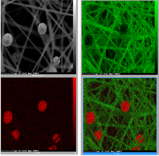


Advanced electron microscopy provides powerful insights into the morphology, microstructure, and surface characteristics of biological specimens, biomaterials, and biomedical devices. Our laboratory offers high-resolution characterization using Field Emission Scanning Electron Microscopy (FE-SEM) combined with Energy Dispersive X-ray Spectroscopy (EDS) to support research, product development, quality assessment, and failure investigations across life science and biotechnology applications.
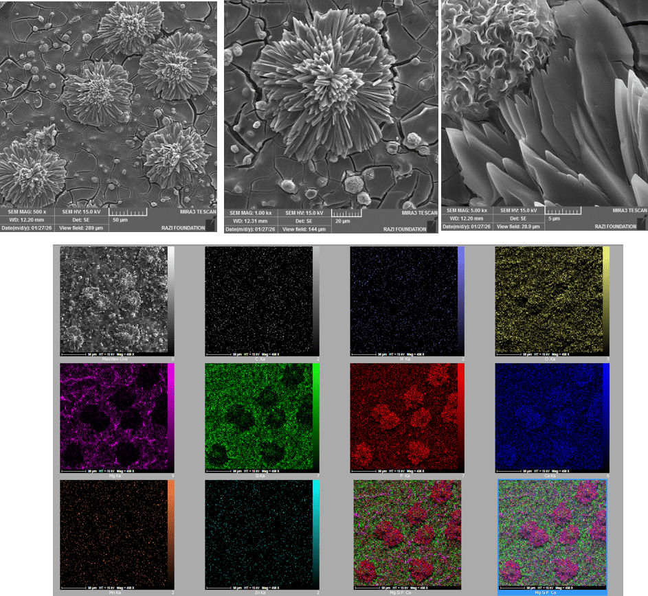
Our analytical capabilities enable detailed examination of biological and engineered materials at the micro- and nanoscale. FE-SEM imaging reveals surface morphology, particle architecture, porosity, structural integrity, and microstructural features, while EDS analysis provides localized elemental composition and distribution, supporting the evaluation of material composition, surface modifications, coatings, and potential contaminants.
By combining advanced imaging with elemental characterization, we provide valuable information for the development and optimization of biomaterials, medical devices, tissue engineering products, drug delivery systems, and biotechnology innovations, helping researchers and manufacturers better understand material performance, biological interactions, and product quality.
Our Services
- Biomaterials Characterization
High-resolution imaging and elemental analysis of biomaterials to evaluate microstructure, surface morphology, porosity, particle morphology, and compositional uniformity for research and product development.
- BioGlass & Bioactive Materials
Characterization of bioglass systems and bioactive ceramics to investigate surface features, microstructure, elemental composition, and material homogeneity for biomedical applications.
- Cell Morphology Analysis
FE-SEM analysis of cultured cells on biomaterials and medical surfaces to evaluate cell attachment, morphology, spreading, proliferation, and cell–material interactions. High-resolution imaging provides valuable insights into cellular response, surface compatibility, and biomaterial performance for tissue engineering and biomedical research.
- Exosome Characterization
High-resolution FE-SEM characterization of exosomes to evaluate particle morphology, structural integrity, size distribution, aggregation, and surface features at the nanoscale, supporting extracellular vesicle research, biomarker studies, and biotechnology applications.
- Biofilm Characterization
High-resolution FE-SEM imaging of biofilms to evaluate their morphology, architecture, microbial colonization, extracellular polymeric substance (EPS) network, and interactions with biomaterial and medical device surfaces. When required, EDS analysis can be performed to investigate elemental composition and mineral deposits associated with biofilm formation, supporting biomedical, environmental, and industrial research.